
Cell Study Guide: An Overview
This comprehensive guide explores the foundational aspects of cell biology‚ encompassing cell structure‚ types‚ transport‚ division‚ and observation techniques.
It’s designed to aid understanding of life’s fundamental units‚ from unicellular organisms to complex multicellular systems‚ utilizing microscopy.
Cell biology is a captivating scientific field dedicated to unraveling the intricacies of cells – the basic building blocks of all known living organisms. From the simplest bacteria to the complex tissues of animals and plants‚ cells represent the fundamental unit of life‚ exhibiting remarkable diversity in structure and function.
This discipline delves into the study of cellular structure‚ physiology‚ and life cycle‚ exploring how cells grow‚ reproduce‚ respond to stimuli‚ and interact with their environment. Understanding cells is paramount to comprehending the processes that underpin life itself‚ including disease mechanisms and potential therapeutic interventions.
The study of cells encompasses a wide range of techniques‚ from microscopy and molecular biology to genetics and biochemistry. Cell biology is not merely a descriptive science; it seeks to explain the ‘how’ and ‘why’ of cellular processes‚ providing a framework for understanding the complexities of life at its most fundamental level. It’s a constantly evolving field‚ driven by new discoveries and technological advancements.
The Cell Theory
The Cell Theory is a cornerstone of modern biology‚ representing a unifying concept that explains the fundamental nature of life. Developed over centuries through the work of numerous scientists‚ it comprises three core tenets that have profoundly shaped our understanding of the biological world.
Firstly‚ all living organisms are composed of one or more cells. This establishes the cell as the basic structural and functional unit of life. Secondly‚ the cell is the basic unit of structure and organization in organisms. Finally‚ all cells arise from pre-existing cells‚ rejecting the idea of spontaneous generation.
These principles‚ initially proposed by Schleiden‚ Schwann‚ and Virchow‚ have withstood the test of time and continue to guide biological research. The Cell Theory isn’t simply a historical artifact; it provides a foundational framework for investigating everything from embryonic development to disease pathology‚ emphasizing the cell’s central role in all life processes.

Cell Structure: Key Components
Cells possess three main parts: the cell membrane‚ nucleus‚ and cytoplasm. These components work in harmony to maintain life’s processes and cellular function.
The Cell Membrane: Structure and Function
The cell membrane acts as a selective barrier‚ surrounding each cell and meticulously controlling the movement of substances both into and out of its interior. This crucial boundary isn’t simply a wall; it’s a dynamic structure primarily composed of a phospholipid bilayer.
Phospholipids arrange themselves with hydrophilic heads facing outward towards the watery environments‚ and hydrophobic tails shielded inward. Proteins are embedded within this bilayer‚ performing diverse functions like transport‚ signaling‚ and structural support.
This fluid mosaic model describes the membrane’s flexibility and the ability of proteins to move laterally. The membrane’s selective permeability is vital for maintaining cellular homeostasis‚ ensuring the right conditions for life processes. It regulates nutrient intake‚ waste removal‚ and communication with the external environment‚ ultimately defining cellular integrity.
Cytoplasm and Cytosol
The cytoplasm is the gel-like substance filling the interior of a cell‚ encompassing everything between the cell membrane and the nucleus. It’s a bustling hub of activity where many essential cellular processes occur‚ providing a medium for organelles to function.
Within the cytoplasm lies the cytosol‚ a fluid component primarily composed of water‚ salts‚ and various organic molecules. This isn’t just empty space; it’s where numerous metabolic pathways‚ like glycolysis‚ take place‚ providing energy for the cell.
The cytoskeleton‚ a network of long fibers‚ resides within the cytoplasm‚ providing structural support and facilitating cell movement. It’s a dynamic framework constantly remodeling itself to meet the cell’s needs‚ ensuring shape and internal organization. The cytoplasm is truly the cell’s internal environment.
The Nucleus: Control Center of the Cell
The nucleus is often described as the cell’s brain‚ serving as the central repository of genetic information in the form of DNA. Enclosed by a double membrane called the nuclear envelope‚ it carefully safeguards the cell’s genome from damage and regulates access.
Within the nucleus‚ DNA is organized into chromosomes‚ structures that become visible during cell division. The nucleus directs protein synthesis by transcribing DNA into RNA‚ which then travels to the ribosomes.

It also contains the nucleolus‚ a region responsible for ribosome assembly. Essentially‚ the nucleus controls all cellular activities‚ from growth and metabolism to reproduction‚ making it indispensable for cell survival and function. It’s the command center of the entire cellular operation.
Organelles: Specialized Structures
Organelles are specialized subunits within a cell‚ each performing a distinct function essential for overall cell survival and operation. These structures‚ found within the cytoplasm of eukaryotic cells‚ work harmoniously to maintain cellular homeostasis.
Key organelles include mitochondria‚ the powerhouses generating energy through cellular respiration; ribosomes‚ responsible for protein synthesis; and the endoplasmic reticulum (ER)‚ involved in protein and lipid production.
The Golgi apparatus processes and packages proteins‚ while lysosomes break down waste materials. The cytoskeleton provides structural support and facilitates movement. Each organelle’s unique role contributes to the cell’s complex functionality‚ demonstrating a remarkable level of cellular organization and efficiency.
Mitochondria: Powerhouse of the Cell
Mitochondria are often referred to as the “powerhouses” of the cell due to their crucial role in generating adenosine triphosphate (ATP)‚ the primary energy currency of the cell. These double-membrane-bound organelles are found in nearly all eukaryotic cells.
Through a process called cellular respiration‚ mitochondria break down glucose and other fuel molecules to produce ATP‚ providing the energy needed for various cellular processes. Their unique structure‚ featuring inner and outer membranes with folded cristae‚ maximizes surface area for ATP production.
Mitochondria also play roles in calcium signaling‚ programmed cell death (apoptosis)‚ and heat generation. Their functionality is vital for maintaining cellular life and overall organismal health‚ demonstrating their central importance.
Ribosomes: Protein Synthesis
Ribosomes are complex molecular machines responsible for protein synthesis‚ a fundamental process for all living cells. These structures are found in both prokaryotic and eukaryotic cells‚ though their composition differs slightly between the two.
Ribosomes read the genetic code carried by messenger RNA (mRNA) and use this information to assemble amino acids into polypeptide chains‚ which then fold into functional proteins. They consist of two subunits – a large and a small subunit – that come together during translation.
Ribosomes can be found free-floating in the cytoplasm or bound to the endoplasmic reticulum (ER)‚ forming rough ER. Proteins synthesized on free ribosomes generally function within the cytoplasm‚ while those made on bound ribosomes are destined for secretion or membrane integration.
Endoplasmic Reticulum (ER): Smooth and Rough
The Endoplasmic Reticulum (ER) is an extensive network of membranes found throughout eukaryotic cells‚ playing a crucial role in various cellular processes. It exists in two primary forms: rough ER and smooth ER‚ distinguished by their structure and function.
Rough ER is studded with ribosomes‚ giving it a “rough” appearance. These ribosomes are sites of protein synthesis‚ and the rough ER is heavily involved in the production and processing of proteins destined for secretion or membrane incorporation.
Smooth ER lacks ribosomes and is involved in lipid synthesis‚ carbohydrate metabolism‚ and detoxification of drugs and poisons. It also stores calcium ions‚ essential for muscle contraction and other cellular signaling pathways. The ER’s interconnected network facilitates transport within the cell.
Golgi Apparatus: Processing and Packaging
The Golgi apparatus‚ a defining organelle in eukaryotic cells‚ functions as a central processing and packaging center for proteins and lipids synthesized in the endoplasmic reticulum. It appears as a stack of flattened‚ membrane-bound sacs called cisternae.
Proteins and lipids travel through the Golgi‚ undergoing further modification‚ sorting‚ and packaging into vesicles. These vesicles bud off from the Golgi and transport their contents to other destinations within the cell‚ or even outside the cell via exocytosis.
The Golgi also synthesizes certain macromolecules‚ such as polysaccharides. Its structure allows for sequential modification of molecules as they move through its compartments‚ ensuring proper protein folding and function. Essentially‚ it prepares cellular products for their specific roles.
Lysosomes: Cellular Digestion
Lysosomes are membrane-bound organelles functioning as the digestive system of the cell‚ crucial for breaking down cellular waste and debris. They contain powerful hydrolytic enzymes capable of digesting a wide range of biomolecules – proteins‚ lipids‚ carbohydrates‚ and nucleic acids.
These enzymes work best in an acidic environment‚ maintained within the lysosome. Lysosomes participate in autophagy‚ the process of self-eating‚ where damaged or unnecessary cellular components are broken down and recycled. They also play a role in phagocytosis‚ engulfing and digesting foreign materials like bacteria.
Essentially‚ lysosomes are responsible for maintaining cellular cleanliness and providing building blocks for new molecules‚ ensuring optimal cell function and preventing the accumulation of harmful substances.

Cell Types: Prokaryotic vs. Eukaryotic
Cells fall into two categories: prokaryotic and eukaryotic‚ distinguished by their internal structures. Eukaryotes possess a nucleus‚ while prokaryotes do not.
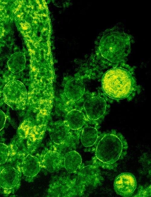
Prokaryotic Cells: Bacteria and Archaea
Prokaryotic cells‚ representing the earliest life forms‚ are characterized by their simple structure and lack of a nucleus. Bacteria and Archaea comprise this domain‚ differing significantly from eukaryotic cells in organization.
These cells typically have a cell wall‚ a cell membrane‚ cytoplasm‚ ribosomes‚ and genetic material in a nucleoid region – but no membrane-bound organelles. Their DNA is usually a single‚ circular chromosome.
Bacteria are ubiquitous‚ found in diverse environments‚ and play crucial roles in ecosystems. Archaea often inhabit extreme environments‚ like hot springs or highly saline waters‚ showcasing unique adaptations.
Prokaryotic cells reproduce primarily through binary fission‚ a simple form of asexual reproduction. This process results in two identical daughter cells. Their smaller size and simpler structure allow for rapid reproduction rates.
Understanding prokaryotic cells is fundamental to comprehending the evolution of life and the diversity of microbial life on Earth‚ impacting fields like medicine and biotechnology.
Eukaryotic Cells: Plants‚ Animals‚ Fungi‚ and Protists
Eukaryotic cells are defined by their complex internal structure‚ most notably the presence of a nucleus – a membrane-bound organelle housing the cell’s DNA. Plants‚ animals‚ fungi‚ and protists are all composed of these cell types.
Unlike prokaryotes‚ eukaryotic cells contain numerous membrane-bound organelles‚ such as mitochondria‚ endoplasmic reticulum‚ and Golgi apparatus‚ each performing specialized functions. This compartmentalization enhances efficiency.
Plant cells possess unique features like cell walls made of cellulose and chloroplasts for photosynthesis. Animal cells lack cell walls and chloroplasts‚ but often have centrioles involved in cell division.
Fungi exhibit cell walls made of chitin‚ while protists are a diverse group‚ some with and some without specific organelles. Eukaryotic cells reproduce through mitosis and meiosis.
Studying eukaryotic cells is crucial for understanding the complexities of multicellular life and the mechanisms driving growth‚ development‚ and disease.

Cell Transport Mechanisms
Cellular transport governs the movement of substances across the cell membrane‚ vital for maintaining homeostasis. This includes both passive and active transport processes.
Passive Transport: Diffusion and Osmosis
Passive transport mechanisms rely on concentration gradients‚ requiring no cellular energy expenditure. Diffusion is the movement of molecules from an area of high concentration to low concentration‚ ultimately achieving equilibrium. This process doesn’t require a membrane‚ but it frequently occurs across it.
Osmosis‚ a specific type of diffusion‚ focuses on the movement of water across a selectively permeable membrane. Water travels from areas of high water concentration (low solute concentration) to areas of low water concentration (high solute concentration).
Factors influencing osmosis include solute concentration and membrane permeability. Understanding these principles is crucial for comprehending cellular behavior in varying environments. Cells can experience hypotonic‚ hypertonic‚ or isotonic conditions‚ impacting water movement and cell volume. These processes are fundamental to nutrient uptake and waste removal in cells.
Active Transport: Requiring Energy
Active transport mechanisms move substances against their concentration gradients‚ necessitating cellular energy‚ typically in the form of ATP. Unlike passive transport‚ this process doesn’t follow the natural flow of molecules.
Key examples include the sodium-potassium pump‚ vital for maintaining cellular membrane potential‚ and endocytosis/exocytosis. Endocytosis involves the cell engulfing substances‚ forming vesicles to bring them inside‚ while exocytosis expels substances by fusing vesicles with the cell membrane.
These processes are essential for nutrient absorption‚ waste removal‚ and nerve impulse transmission. Active transport allows cells to maintain internal conditions different from their surroundings‚ crucial for survival. Carrier proteins facilitate the movement of specific molecules‚ requiring ATP binding and conformational changes. Understanding active transport is key to understanding cellular function.

Cell Division and the Cell Cycle
Cell division‚ encompassing mitosis and meiosis‚ is fundamental for growth‚ repair‚ and reproduction. The cell cycle regulates these processes‚ ensuring genetic integrity.
Mitosis: Cell Replication
Mitosis is a fundamental process of cell division resulting in two identical daughter cells from a single parent cell. This process is crucial for growth‚ tissue repair‚ and asexual reproduction in organisms.
The mitotic phase is characterized by distinct stages: prophase‚ metaphase‚ anaphase‚ and telophase. During prophase‚ chromosomes condense and become visible‚ while the nuclear envelope breaks down. Metaphase involves the alignment of chromosomes along the cell’s equator.
Anaphase sees the separation of sister chromatids‚ pulled towards opposite poles of the cell. Finally‚ telophase culminates in the formation of two new nuclei‚ each containing a complete set of chromosomes.
Cytokinesis‚ often occurring concurrently with telophase‚ physically divides the cytoplasm‚ resulting in two separate and identical daughter cells. Accurate chromosome segregation during mitosis is vital to maintain genetic stability and prevent errors that could lead to cellular dysfunction or disease.
Meiosis: Sexual Reproduction
Meiosis is a specialized cell division process essential for sexual reproduction in eukaryotic organisms. Unlike mitosis‚ which produces identical cells‚ meiosis generates genetically diverse gametes – sperm and egg cells – with half the number of chromosomes as the parent cell.
This reduction in chromosome number is achieved through two rounds of division: Meiosis I and Meiosis II. Meiosis I involves the separation of homologous chromosome pairs‚ leading to genetic recombination through crossing over‚ increasing genetic diversity.
Meiosis II resembles mitosis‚ separating sister chromatids. The resulting four haploid daughter cells‚ each containing a unique combination of genetic material‚ are gametes ready for fertilization.
Fertilization restores the diploid chromosome number‚ creating a zygote with a blend of genetic information from both parents. Meiosis ensures genetic variation within populations‚ driving evolution and adaptation.

Microscopy and Cell Observation
Microscopy is crucial for visualizing cells and their components‚ enabling detailed study of cellular structures. Various techniques and sample preparation methods are utilized.
Types of Microscopes
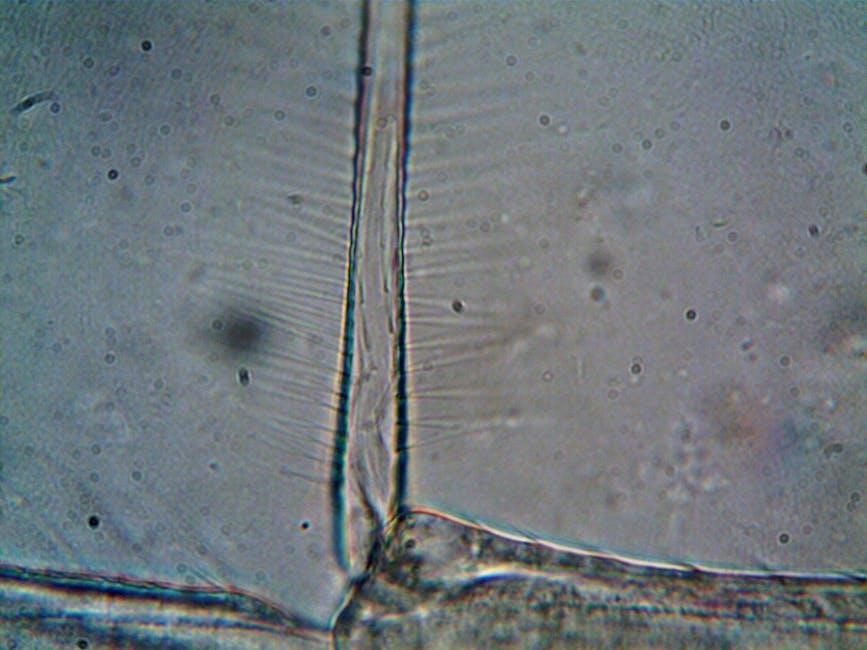
Several types of microscopes are employed in cell observation‚ each offering unique capabilities. Light microscopes‚ the most common type‚ use lenses to magnify images using visible light‚ allowing for the observation of stained or naturally pigmented cells. These are relatively inexpensive and easy to use.
However‚ resolution is limited by the wavelength of light. Electron microscopes‚ utilizing beams of electrons instead of light‚ achieve significantly higher magnification and resolution‚ revealing ultrastructural details. Transmission Electron Microscopy (TEM) examines thin sections of cells‚ while Scanning Electron Microscopy (SEM) provides detailed surface views.
Confocal microscopy creates sharp‚ three-dimensional images by scanning a specimen with a laser beam. Fluorescence microscopy uses fluorescent dyes to label specific cellular components‚ enabling their visualization. Phase-contrast microscopy enhances contrast in transparent specimens‚ and dark-field microscopy illuminates specimens against a dark background. The choice of microscope depends on the specific research question and the level of detail required.
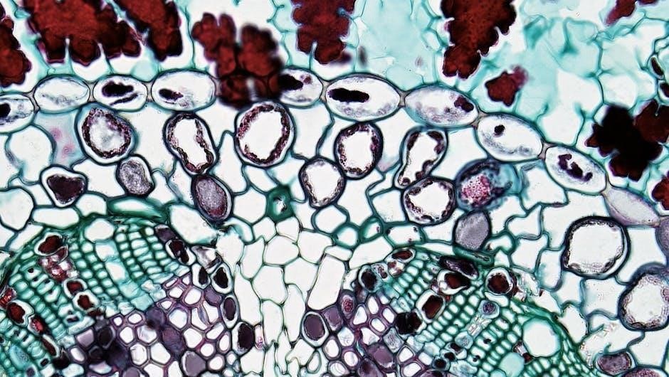
Preparing Cell Samples for Observation
Proper sample preparation is crucial for effective cell observation under a microscope. For light microscopy‚ samples are often stained with dyes to enhance contrast and highlight specific structures. Common stains include methylene blue and eosin. Samples may be mounted on slides with a coverslip to flatten them and provide a protective layer.
For electron microscopy‚ more extensive preparation is required. Cells are typically fixed to preserve their structure‚ then embedded in resin and sliced into ultra-thin sections using an ultramicrotome. These sections are stained with heavy metals to provide contrast.
Live cell imaging requires maintaining cell viability‚ often using specialized chambers and media. Techniques like immunofluorescence involve labeling specific proteins with fluorescent antibodies. Careful attention to detail during preparation minimizes artifacts and ensures accurate visualization of cellular components‚ leading to reliable results.